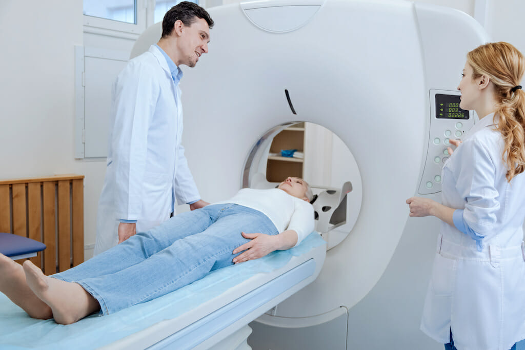
A computerized tomography (CT) scan allows radiologists to capture x-ray images of the human anatomy to help examine abnormalities and diagnose certain conditions or injuries that affect our organs, bones, muscles, and blood vessels [1].
Unlike a standard x-ray image, CT scans utilize computerized technology to take multiple images of the body from different angles, which it then puts together to show a detailed cross-sectional slice of the area [2].
Clinicians will request patients to undergo CT scans for various reasons, and a patient may have additional tests performed before or after the procedure is completed.
For example, in the case of lung disease detection, your healthcare team may recommend an initial chest x-ray to inspect the lungs for damage before having you undergo a CT scan. Chest x-rays can typically show specific abnormalities that require further investigation via CT scans [3].
Using CT Scans for COPD Detection
A clinician may recommend a CT scan if you’re experiencing symptoms of emphysema—a type of chronic obstructive pulmonary disease (COPD) where a person’s alveoli (i.e., air sacs) become irreversibly damaged and stretched.
As with other lung conditions that restrict the airways, emphysema can make breathing and distributing oxygen throughout the bloodstream difficult. However, there are three different types of emphysema that affect different parts of the lungs.
 These are known as:
- Paraseptal emphysema
- Centrilobular emphysema
- Panlobular emphysema [4]
Your clinician may recommend a CT scan to help diagnose which of the three conditions you may be experiencing.
Read our article on emphysema for more information on this condition.
FAQs about CT Scans for the Lungs
In honor of COPD Awareness Month, we are addressing common questions you or your loved ones may have regarding CT scans.
![]()
What Can a CT Scan of the Lungs Detect?
As mentioned, CT scans can help clinicians diagnose certain conditions affecting different bodily organs. They also help guide professionals in conducting medical procedures, including biopsies and surgeries [5].
In the case of lung disease detection, CT scans can help pinpoint the location of an infection or inflammation that may be damaging the lungs and caused by an underlying condition, such as COPD.

How Long Does a CT Scan Take for Lungs?
On average, CT scans can take up to an hour or longer. The procedure itself may only take up to 30 minutes [6], but this will depend on each individual situation.
You may also receive the results within 24 hours of the test. Patients can usually resume their regular activities following the procedure—though your clinician may give you medication to help you relax during the scan, which may affect your ability to drive home afterward.
What Is Considered an Abnormal CT Scan of the Lungs?
As noted earlier, your clinician may recommend a CT scan following a chest x-ray that shows abnormalities. The CT scan allows your clinician to capture a 2-D image of your lungs’ structure to investigate these abnormalities more closely.
Using a CT scan, your clinician can also check for signs of lung damage, infection, or airway obstruction. Sometimes, a clinician may use a contrast agent (or dye) during a CT scan to help improve the images and highlight specific features of the lungs [7].

How Does a CT Scan Differ from a High-Resolution CT Scan?
As you might imagine, high-resolution CT scans (or HRCT) help clinicians create more detailed images of the lungs using higher-resolution technology. HRCT scans allow clinicians to create 3-D images of the lungs’ structure, allowing them to detect underlying conditions that may be causing symptoms like shortness of breath, chronic cough, and lung infections to develop.
CT scans can help diagnose various health conditions and injuries, but only an HRCT scan can diagnose bronchiectasis. Bronchiectasis is a chronic lung condition that damages the airways in the lungs from repeated inflammation and infection, causing them to widen.
HRCT scans allow clinicians to identify the damage caused by bronchiectasis or the prevalence of its symptoms, especially in patients with COPD.

Find More COPD Resources
Undergoing a CT scan for the first time can feel daunting. Therefore, we hope this article clears up any misgivings or confusion on how this technology works, so you feel confident going into the procedure and sharing this information with others in your community.
For more information on COPD or bronchiectasis, stay connected to our blog page for insights, research, and helpful articles!

Resources
[1] Johns Hopkins Medicine. “Computerized Tomography (CT) Scan.” Retrieved from https://www.hopkinsmedicine.org/health/treatment-tests-and-therapies/computed-tomography-ct-scan
[2] Cleveland Clinic. “CT (Computerized Tomography) Scan.” Retrieved from https://my.clevelandclinic.org/health/diagnostics/4808-ct-computed-tomography-scan
[3] American Lung Association. “What Is a CT Scan?” Retrieved from https://www.lung.org/lung-health-diseases/lung-procedures-and-tests/ct-scan
[4] Medical News Today. “What Is Emphysema?” Retrieved from https://www.medicalnewstoday.com/articles/8934
[5] Mayo Clinic. “CT Scan: Overview.” Retrieved from https://www.mayoclinic.org/tests-procedures/ct-scan/about/pac-20393675
[6] Cleveland Clinic. “CT (Computerized Tomography) Scan.” Retrieved from https://my.clevelandclinic.org/health/diagnostics/4808-ct-computed-tomography-scan
[7] Cleveland Clinic. “CT (Computerized Tomography) Scan.” Retrieved from https://my.clevelandclinic.org/health/diagnostics/4808-ct-computed-tomography-scan