
High-resolution computerized technology (HRCT) allows clinicians to get an accurate bronchiectasis diagnosis among patients experiencing common symptoms of this condition.
Bronchiectasis (bron-kee-eck-tuh-sis) is an irreversible chronic lung condition caused by abnormal widening of the lung’s airways. Although no cure for bronchiectasis exists, patients can manage their symptoms and avoid triggering flare-ups that may require hospitalizations.
What Is HRCT Scanning?
High-resolution CT scanning is a medical imaging technique that uses X-rays to create detailed images of the body’s internal structures. It provides a clear view of your body’s bones, blood vessels, and organs, allowing doctors to diagnose and treat a wide range of conditions.¹
 HRCT scans are often used to detect lung cancer, pulmonary fibrosis, and other respiratory conditions. In fact, the technology creates 3D models of your lungs by combining images to show their size, shape, and position.²
Additionally, these models help determine the cause of lung symptoms, such as shortness of breath, or check for lung problems.³
CT vs. HRCT? What’s the Difference?
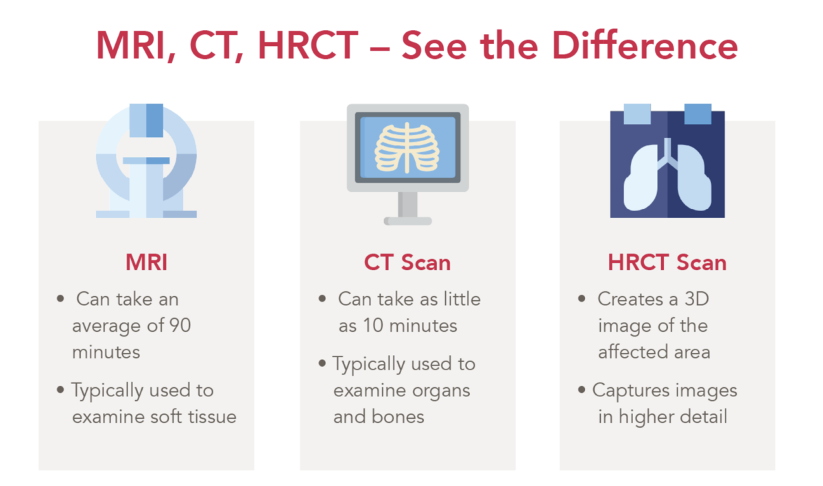
CT stands for computerized (or computed) tomography. The main difference between a CT vs. an HRCT scan is the level of detail they provide.
HRCT uses thinner slices (i.e., detailed views) at higher resolutions to produce 3D models, making it particularly useful for examining small structures like the lungs’ airways.4
Remember, bronchiectasis is categorized as an abnormal “widening” of the airways; therefore, other diagnostic tools may not be appropriate for detecting early signs of this chronic condition.
However, CT scans are still incredibly helpful in diagnosing other lung conditions like COPD (chronic obstructive pulmonary disease).
Your doctor will determine which type of scan is best for your situation.
HRCT vs. MRI? How Do They Differ?
An MRI, which stands for Magnetic Resonance Imaging, is a type of medical imaging that uses a strong magnetic field and radio waves to create detailed images of the inside of the body.5
MRI scans are commonly used to examine soft tissue and diagnose various conditions, including cartilage loss, torn ligaments, spine injury, or joint inflammation.6
Unlike X-rays and HRCT scans, MRI does not use ionizing radiation; however, it typically does not produce the same level of detail that would benefit diagnosing a condition like bronchiectasis.
Additionally, MRIs can also take 90 minutes,7 depending on the area being examined, while CT scans can take as little as 10 minutes.8
HRCT for Chest Imaging: What Are the Benefits?
In our video series “Ask a Pulmonologist,” featuring Dr. Frederic Seifer—a renowned pulmonologist—Dr. Seifer discusses the importance of getting an HRCT scan, especially for patients who may be experiencing a comorbid condition of COPD and bronchiectasis.
According to Dr. Seifer, an HRCT scan is an effective way to ensure clinicians adequately diagnose a prevalence of bronchiectasis and prescribe the right treatment plan to help patients manage their symptoms.
What to Expect During an HRCT Scan
If you request, or your doctor recommends, an HRCT scan to evaluate your lungs for signs of bronchiectasis, you will be placed lying down on your back, on top of a table that slides into a tunnel-like scanner.9
- You will be asked to stay as still as possible.
- From inside the tunnel, the scanner will begin taking multiple images of your chest and lungs to create a computerized 3D image of the chest wall.
- Your clinician may also ask you to hold your breath for a short period of time, usually fewer than 15 to 20 seconds.
- The table moves back out of the scanner when the exam is over.
Again, this procedure takes as little as 10 minutes and is painless.10

What to Expect Following an HRCT Scan
If your clinician determines that your symptoms (i.e., shortness of breath, frequent cough, recurring lower respiratory infections, etc.), are a sign of bronchiectasis, the next step is prescribing an effective airway clearance device to help you manage symptoms and find relief.
The SmartVest Airway Clearance System is a comfortable11 therapy device that uses high-frequency chest wall oscillation (HFCWO) to deliver 360° chest coverage. SmartVest helps your lungs propel mucus up and out of the airways, reducing the risk of recurring infections and lung damage.12
To learn more about how it works, you can schedule a chat with one of our patient care advocates or request an information packet to start your journey to a lifetime of airway clearance.

Resources
[1] National Heart, Lung, and Blood Institute. “Chest CT Scan.” Retrieved from https://www.nhlbi.nih.gov/health-topics/chest-ct-scan
[2] National Library of Medicine. “High-resolution CT of the lungs: Indications and diagnosis.” Retrieved from https://pubmed.ncbi.nlm.nih.gov/29243467/
[3] National Heart, Lung, and Blood Institute. “Chest CT Scan.” Retrieved from https://www.nhlbi.nih.gov/health-topics/chest-ct-scan
[4] National Heart, Lung, and Blood Institute. “Chest CT Scan.” Retrieved from https://www.nhlbi.nih.gov/health-topics/chest-ct-scan
[5] National Institute of Biomedical Imaging and Bioengineering. “Magnetic Resonance Imaging (MRI): Retried from https://www.nibib.nih.gov/science-education/science-topics/magnetic-resonance-imaging-mri
[6] Johns Hopkins Medicine. “CT Scan Versus MRI Versus X-Ray: What Type of Imaging Do I Need?” Retrieved from https://www.hopkinsmedicine.org/health/treatment-tests-and-therapies/ct-vs-mri-vs-xray
[7] NHS. “MRI Scans: How It’s Performed.” Retrieved from https://www.nhs.uk/conditions/mri-scan/what-happens/
[8] Cleveland Clinic. “CT (Computed Tomography) Scan.” Retrieved from https://my.clevelandclinic.org/health/diagnostics/4808-ct-computed-tomography-scan
[9] Cleveland Clinic. “CT (Computed Tomography) Scan.” Retrieved from https://my.clevelandclinic.org/health/diagnostics/4808-ct-computed-tomography-scan
[10] Cleveland Clinic. “CT (Computed Tomography) Scan.” Retrieved from https://my.clevelandclinic.org/health/diagnostics/4808-ct-computed-tomography-scan
[11] Pokorney J. Comparison of Oscillatory Trough Pressure Generated by High Frequency Chest Wall Oscillation (HFCWO) Systems: A White Paper.
[12] Siefert, C. et al. Respiratory Therapy, Vol. 11 No 4, 34-38, 2016.